
Showing posts with label Pediatrics. Show all posts
Showing posts with label Pediatrics. Show all posts
Tuesday, February 14, 2012

Friday, June 17, 2011

Friday, February 11, 2011
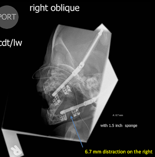
New device for mandibular distraction
Dr Grant Fairbanks is using a new device for mandibular distraction.
It is threaded but not ratcheted and therefore can unscrew, losing ground.
He will be obtaining followup radiographs to check the distraction every 2-3 weeks on patients and would like an AP like the image attached as well as bilateral obliques with the interested side flat against the cassette to minimize magnification and obtain a reproducible measurement. The obliques may need a little cephalic angulation to prevent superimposition of the hardware. He is interested in the distance between the two plates. Their inner margins mark the edges of the osteotomy.
Attached is a sample Powerscribe macro if you like.
It is threaded but not ratcheted and therefore can unscrew, losing ground.
He will be obtaining followup radiographs to check the distraction every 2-3 weeks on patients and would like an AP like the image attached as well as bilateral obliques with the interested side flat against the cassette to minimize magnification and obtain a reproducible measurement. The obliques may need a little cephalic angulation to prevent superimposition of the hardware. He is interested in the distance between the two plates. Their inner margins mark the edges of the osteotomy.
Attached is a sample Powerscribe macro if you like.
[] []
Clinical History: [<Pierre Robin syndrome, micrognathia, mandible distractors>]
Findings: Status post bilateral mandibular distraction. [<An enteric tube is present.>] [<There is no evidence of breakage of the hardware or loosening of the screws.>]
Distraction
Left: [] mm
Right: [] mm.
Impression: [<There is no evidence of loosening or breakage of the hardware.>]
Code [<20>]

Clinical History: [<Pierre Robin syndrome, micrognathia, mandible distractors>]
Findings: Status post bilateral mandibular distraction. [<An enteric tube is present.>] [<There is no evidence of breakage of the hardware or loosening of the screws.>]
Distraction
Left: [] mm
Right: [] mm.
Impression: [<There is no evidence of loosening or breakage of the hardware.>]
Code [<20>]


Thanks to Bill Andolsek, DO
Wednesday, January 19, 2011
Case of the Week: 20 month old male with a specific bone finding


Thursday, December 30, 2010
Sedation Services for Medical Imaging - baskinhj@gmail.com
Sedation Nurse Practitioner Services for Medical Imaging
In House Hours: (physically present in Medical Imaging)
Mon-Fri: 0630-1700
Sat: 07:30-1300
On-Call Hours:
Sat: 1300-1600
Sun: 07:30-1600
To schedule sedation during On-Call hours:
1. Page the Sedation NP on call: 801-339-6578
1. Page the Sedation NP on call: 801-339-6578
2. NP will get the patient info from the radiologist and will call and consult with ordering physician. NP will assess via telephone if patient is an appropriate candidate for sedation. If not, NP will tell the ordering physician that they should consult anesthesia for other sedation options.
3. If NP feels patient is a sedation candidate, the NP will call and coordinate an imaging start time between the MRI/nuc med technologist and the radiology sedation nurse. NP will triage sedation imaging studies along with hospital wide sedation needs.
4. Sedation recovery will be provided by radiology sedation RN. NP will be available by pager following imaging study for any additional sedation recovery issues.
5. Radiologists will notify the sedation NP as soon as the request for a sedated procedure has been received
6. PICU and NICU patients needing MRI studies afterhours and weekends generally provide their own sedation. Generally, this is adequate sedation
GA Sedation for Urgent/Emergent MRI studies:
- A few Holidays during the year. Page sedation NP to determine if they are working on a specific holiday.
- weekdays between 1700 and overnight
- weekends between 1600 and overnight
- Ordering physician will have to consult for GA sedation (Via smartweb, hospital operator, or OR front desk 662-2750) Since radiologist does not know the clinical details of a patient, it is most appropriate that the decision to use GA for sedation comes from the ordering physician
- Have MRI tech coordinate imaging time with anesthesiologist
- Radiology sedation RN are not involved with GA sedation
- This GA sedation protocol for urgent imaging studies should include ER patients requiring imaging during these hours
Friday, December 3, 2010
Case of the Week: Hard left supraclavicular mass


Monday, November 29, 2010
Liver volumes

Spleen volumes

Wednesday, November 24, 2010
Femoral/tibial torsion studies
,


Regarding Femoral/tibial torsion studies:
Dr. Stevens would like us to:
Dr. Stevens would like us to:
1) Use the screen layout that I show in the second and third attachments
2) Save our measurements when closing the study
3) Use the macro below
Also, please read the note on the first attachment regarding how he'd like us to measure the femoral neck.
PS: The line about P. STEVENS in the macro is because Aoki does it differently.
_____________________________
Examination: [<Right>] MRI lower extremity without contrast limited. [] hours.
Examination: [<Right>] MRI lower extremity without contrast limited. [] hours.
Comparison: Contralateral side.
History: Femoral anteversion. [<Lower extremity torsion.>]
Technique: Oblique PD axial images were obtained through the [left/right] hip. Native axial images were obtained through the hip, knee, and ankle.
Findings:
No fracture, periosteal reaction, or other acute osseous abnormality is present in the visualized portions of the lower extremities. Soft tissues are normal appearing.
BY THE METHOD OF P. STEVENS AND B. NIXON,
lower extremity anteversion and torsion angles are calculated as listed below:
Impression: Femoral [<anteversion >] is [] degrees. There is [] degrees femoral [<internal>] torsion and [] degrees tibial [<external>] torsion. Please see separate dictation of the contralateral limb.
_____________________________
Tuesday, November 23, 2010
Case of the week: 21 month old with stridor


The ingestion of disk batteries is particularly important because saliva acts as an electrolyte bath, causing batteries to corrode and leak a solution of sodium or potassium hydroxide. This caustic solution causes rapid destruction of the esophageal wall, extensive edema, and narrowing of the adjacent airway.
Saturday, November 20, 2010
Head US and assessment of gestational age
Sagittal US views
Superior temporal sulcus (STS) 23 weeks
Saw tooth serrations STS (secondary sulcation) 35 weeks
Cingulate sulcus (CS) 29-30 weeks
Marginal branch of CS 32 weeks
Long and short insular gyri 39-40 weeks
Left 40 weeks
Right 39 weeks
Coronal US views
Superior frontal sulcus (SFS) – primary sulcation 25 weeks
Secondary sulcation of SFS 27 weeks
Adapted by Kevin Moore
Thursday, November 18, 2010
Case of the week: 29 month old with elbow pain

Wednesday, November 10, 2010
Google Chrome links
Extensions
Allow you to change the way the browser looks (all black for radiologists!)
- GMail checker: Adds a pop-up feature for checking/deleating/marking spam on your gmail account
- EI Tab: Allows you to stay within Chrome's tabs and emulate Internet Explorer
- Stylish: Allows you to change a site's appearance by adding styles available HERE
Allow you to change the way the browser looks (all black for radiologists!)

Primary Children's On-Line Resources
Access PCMC on-line resources!
You need to use IE or Chrome with THIS EI TAB EXTENSION:
Chrome Version: ER Patient Tracker
Chrome Version: HELP2
You need to use IE or Chrome with THIS EI TAB EXTENSION:
Chrome Version: ER Patient Tracker
Chrome Version: HELP2
If you need to use Internet Explorer, then use these links:
Tuesday, November 9, 2010
Bone age determination in infants

Findings: Using the method described by Elgenmark (Acta Paediat. 33:1, 1946), a child aged [<>] months should have [<>] ossified growth centers. This child has [<>], [<within 2 standard deviations of mean.>]
Impression: [<Normal bone age.>]
Code 10

Sunday, November 7, 2010
Risser Classification

At about 14 years, ossification begins on the anterolateral aspect of the iliac crest and continues posteromedially.
Fusion of the ossified apophysis begins around the age of 15 years and may not be complete until 25 years.
Saturday, November 6, 2010
Skeletal dysplasia: An update
The skeletal dysostoses and dysplasias are now classified as "Genetic Skeletal Disorders," a nod towards our better understanding of the human genome and the genetic basis of disease. Check out the new standard for classifying these disorders online at:
http://www.isds.ch/Nosology2006.html
http://www.isds.ch/Nosology2006.html
Subscribe to:
Posts (Atom)